The Art of the Central Nervous System
A microscopy art page dedicated to celebrating the brain's intricate and captivating beauty and making neuroscience more engaging and accessible to a broader audience.
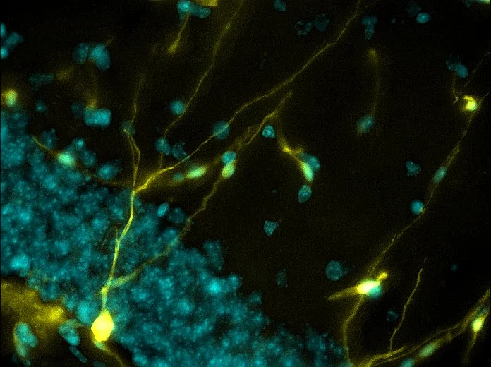
The Brain's Boundless Growth: A neuron (yellow) is genetically labeled with a stem cell marker in the mouse hippocampus. This labeling signifies neurogenesis (the birth of new neurons) and illustrates the brain's capacity for growth.
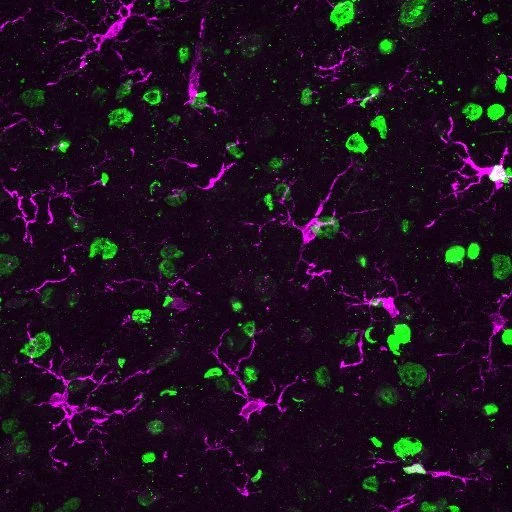
A Symphony of Innate Immune Responses: After spinal cord injury, innate immune cells (magenta) colocalize with phosphatidylserine (green), a fat molecule externalized to the neuron membrane. This colocalization suggests an ongoing response to the injury, indicating that phosphatidylserine is active in regulating inflammation.
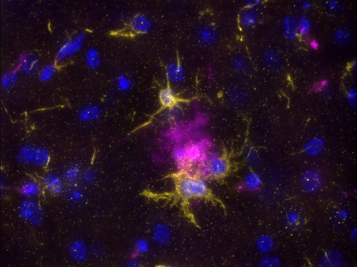
Guardians of Memory: Microglia (yellow), immune cells residing in the brain, are actively engulfing and eliminating the buildup of Alzheimer's Disease plaques (magenta). This image illustrates the brain's innate defense mechanisms and highlights microglia as a potential target for therapeutic intervention.
Complementing Degeneration After Spinal Cord Injury: Complement protein C1q (green) is expressed on degenerating motor neurons (magenta) after spinal cord injury. The complex relationship between inflammation and neural damage provides valuable insights for potential injury treatment.
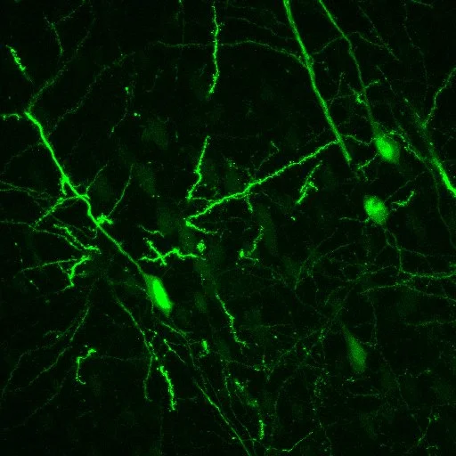
Tracing the Blueprint of Movement: Mouse cortical neurons have been retrogradely traced (viral labeling) from the spinal cord. This labeling technique provides insights into neural circuitry, contributing to understanding motor control mechanisms.
Where Forelimb Control Pathways Merge: The convergence of neural pathways is labeled from the brain to the spinal cord (red, anterograde virus) and spinal cord to brain labeling (green, retrograde virus.) This technique highlights a specific group of brain stem neurons (green & red) that play a crucial role in forelimb motor control.
Microglia in Motion: In a stroke mouse model, microglia (green) are reactive. Genetically labeled blood vessels and astrocytes are shown in magenta using a stem cell marker. The interplay of cellular responses and potential regenerative processes following a stroke provides valuable insights into potential therapeutic approaches.
Plaque Your Brain: This montage displays a mouse model of Alzheimer's Disease, featuring amyloid beta plaques (bright cyan spots) distributed across multiple brain regions. This image illustrates the effect of the disease on different structures of the brain.
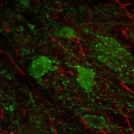
Synaptic Conversations: In the spinal cord, tiny excitatory connections called glutamatergic synapses (red) communicate with motor neurons (green). Our muscles are controlled by the spinal cord through a network of connections between synapses and neurons, which work together like phone lines.
The Forelimb Maestros: Neurons labeled in magenta (anterograde virus tracing originating from the brain stem) and cyan (retrograde viral tracing from the spinal cord) highlight a specific neuron population dedicated to forelimb function. This dual labeling approach offers a glimpse of the neural circuitry within the brain stem responsible for controlling forelimb movements.


